
Heart failure remains a leading cause of morbidity worldwide, and the emergence of continuous wearable electrocardiogram (ECG) technology offers a new frontier for early detection and disease monitoring. In 2026, regulatory bodies such as the FDA and EMA have tightened requirements for digital biomarker validation, demanding robust evidence of clinical relevance, data integrity, and algorithmic transparency. This guide walks you through a systematic, step‑by‑step process for leveraging wearable ECG data to build, validate, and submit a heart‑failure digital biomarker that meets these stringent standards.
1. Define the Digital Biomarker Landscape for Heart Failure
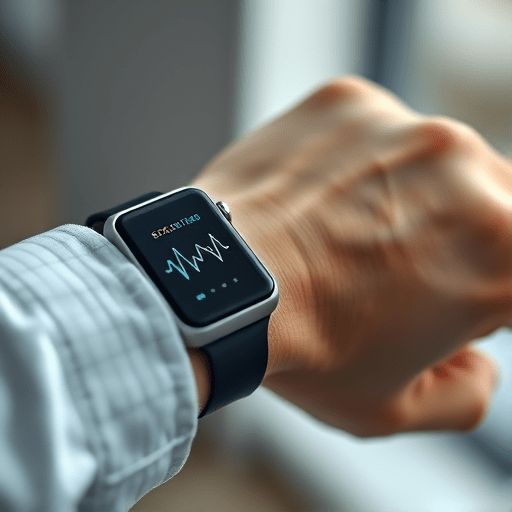
A digital biomarker is a quantifiable, objective measurement derived from digital devices that reflects a biological or clinical state. For heart failure, relevant biomarkers include arrhythmia burden, heart rate variability, conduction delays, and heart rate turbulence. Before you start collecting ECG data, clarify the clinical endpoint your biomarker will address—whether it is risk stratification, early decompensation detection, or therapeutic response monitoring.
1.1 Regulatory Expectations and 2026 Guidance
The 2026 FDA Digital Health Device Guidance emphasizes a “data‑driven” validation pathway. Key expectations include:
- Provenance and lineage: Every raw ECG sample must be traceable to its source device, timestamp, and acquisition settings.
- Clinical relevance: The biomarker must correlate with accepted heart‑failure outcomes (e.g., hospitalization, mortality).
- Algorithm transparency: The processing pipeline must be fully documented, including preprocessing, feature extraction, and decision thresholds.
- Post‑market surveillance: Real‑world evidence (RWE) is required to confirm ongoing safety and effectiveness.
1.2 Clinical Endpoints and Data Needs
Identify the clinical outcomes you will use for validation. Common endpoints include:
- Hospital admission for heart failure exacerbation
- All‑cause mortality within 12 months
- Composite endpoints of rehospitalization or death
Define the statistical power you need to detect clinically meaningful differences. This will drive your sample size calculations and the duration of ECG monitoring required.
2. Selecting the Wearable ECG Platform
Choosing the right device is foundational. In 2026, many consumer wearables have achieved medical‑grade certification, but you still need to assess each product against your validation criteria.
2.1 Signal Fidelity and Battery Life
Look for devices that provide 12‑lead or at least 3‑lead ECG recordings with a sampling rate ≥200 Hz. Signal‑to‑noise ratio (SNR) should be ≥15 dB, and artifact rejection algorithms must be documented. Battery life is critical for continuous monitoring; aim for ≥14 days of operation with automatic data upload when connected to Wi‑Fi.
2.2 Data Security and Interoperability
All data must be encrypted in transit (TLS 1.3 or higher) and at rest (AES‑256). Devices should support standard data formats such as HL7 FHIR or DICOM‑ECG for seamless integration into electronic health records (EHRs). Verify that the manufacturer provides an API for automated data extraction, and that audit trails are maintained for each upload.
3. Designing the Continuous ECG Data Collection Protocol
Protocol design should align with regulatory expectations while ensuring data quality. The following sub‑steps will guide you from cohort selection to feature extraction.
3.1 Cohort Selection and Sample Size Calculations
Recruit a diverse cohort reflecting real‑world heart‑failure populations—age, sex, comorbidities, and socioeconomic status should mirror the target demographic. Use power analysis tools (e.g., G*Power) to estimate the required number of participants. For a 10% event rate and a hazard ratio of 0.75, a sample of 1,200 patients monitored for 12 months typically yields >80% power.
3.2 Signal Preprocessing and Feature Extraction
Preprocessing pipelines should include:
- Baseline wander removal: High‑pass filter at 0.5 Hz.
- Power‑line interference rejection: Notch filter at 50/60 Hz.
- R‑peak detection: Pan‑Tompkins algorithm with adaptive thresholding.
- Artifact detection: Machine‑learning model trained on labeled noise events.
From the cleaned signal, extract features that are predictive of heart‑failure status:
- Heart rate variability (SDNN, RMSSD)
- QT interval dispersion
- Non‑linear dynamics (sample entropy)
- Arrhythmia burden (SVT, AF episodes)
4. Statistical Validation Framework
Validation must demonstrate that the biomarker reliably predicts clinical outcomes across diverse settings. Adopt a two‑stage approach: internal validation with the cohort, followed by external validation with an independent dataset.
4.1 Longitudinal Correlation with Clinical Outcomes
Use Cox proportional hazards models to relate biomarker values to time‑to‑event endpoints. Adjust for covariates such as age, sex, and baseline ejection fraction. Report hazard ratios, confidence intervals, and p‑values. Additionally, calculate the concordance index (C‑index) to assess predictive discrimination.
4.2 ROC and Decision‑Curve Analysis
Generate receiver operating characteristic (ROC) curves to determine optimal cut‑off points. Report area under the curve (AUC) with bootstrapped confidence intervals. Decision‑curve analysis (DCA) will help quantify the net clinical benefit across a range of threshold probabilities, guiding how the biomarker might be integrated into clinical decision support.
5. Integrating with Regulatory Submissions
When you move from validation to submission, meticulous documentation is critical. The FDA’s Digital Health Device Guidance (2026) provides a template for the “Software as a Medical Device” (SaMD) dossier.
5.1 Documenting Data Provenance
For each ECG file, include:
- Device serial number and firmware version
- Acquisition timestamp (UTC) and time zone offset
- Signal quality metrics (SNR, artifact percentage)
- Processing pipeline version
Store this metadata in a relational database with immutable audit logs. This ensures traceability required by the FDA’s “Data Integrity” guidelines.
5.2 Using the 2026 FDA Digital Health Device Guidance
In your submission, align the following sections with the guidance:
- Device Description and Intended Use
- Clinical Validation Plan and Results
- Risk Analysis (FMEA, QRA)
- Post‑Market Surveillance Strategy
- Cybersecurity Plan (ISO 27001 compliance)
Include a concise summary of the biomarker’s performance metrics, a risk‑benefit assessment, and a plan for periodic updates (e.g., algorithm retraining).
6. Post‑Launch Monitoring and Continuous Improvement
Once the biomarker is in clinical use, continuous data collection will feed back into refinement and regulatory compliance.
6.1 Real‑World Evidence and Adaptive Iteration
Aggregate RWE from diverse sites to detect shifts in performance. Employ adaptive trial designs where thresholds can be recalibrated based on ongoing data, ensuring sustained clinical relevance.
6.2 Feedback Loops to Biomarker Algorithms
Establish a closed‑loop system: flagged events trigger clinician review; clinician annotations feed into a supervised learning model to improve detection accuracy. Document all algorithm updates and maintain version control.
By following this comprehensive, step‑by‑step framework, researchers and developers can translate continuous wearable ECG data into a regulatory‑approved digital biomarker that reliably informs heart‑failure management in 2026 and beyond.